| The Perth Group
The HIV-AIDS debate | 
|
| The Perth Group
The HIV-AIDS debate |
|
 Home What the Perth Group has argued
Papadopulos redox theory of cellular function papers Papers and letters published in scientific journals Monograph on mother-to-child transmission
Papers published in Continuum magazine
Papers published in the popular press
Papers/letters rejected by the scientific press
Presentations
Interviews
Selected email correspondence
A virus like no other
Oxidation, Montagnier and the Perth Group Home What the Perth Group has argued
Papadopulos redox theory of cellular function papers Papers and letters published in scientific journals Monograph on mother-to-child transmission
Papers published in Continuum magazine
Papers published in the popular press
Papers/letters rejected by the scientific press
Presentations
Interviews
Selected email correspondence
A virus like no other
Oxidation, Montagnier and the Perth Group
 Montagnier Nobel Prize 2008
The Parenzee Case
The House of Numbers
Latest files
National Libary of Australia Intervew 1993
Others
Africa/South Africa
Questions and answers
Response to the NIH "Evidence" that HIV causes AIDS
Translations of the Perth Group papers
BMJ Online Debate
Contact Us
About the Perth Group
Perth Group at Virusmyth
The Perth Group on YouTube Montagnier Nobel Prize 2008
The Parenzee Case
The House of Numbers
Latest files
National Libary of Australia Intervew 1993
Others
Africa/South Africa
Questions and answers
Response to the NIH "Evidence" that HIV causes AIDS
Translations of the Perth Group papers
BMJ Online Debate
Contact Us
About the Perth Group
Perth Group at Virusmyth
The Perth Group on YouTube
|
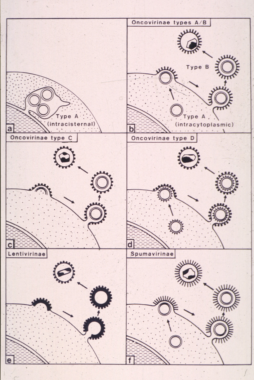
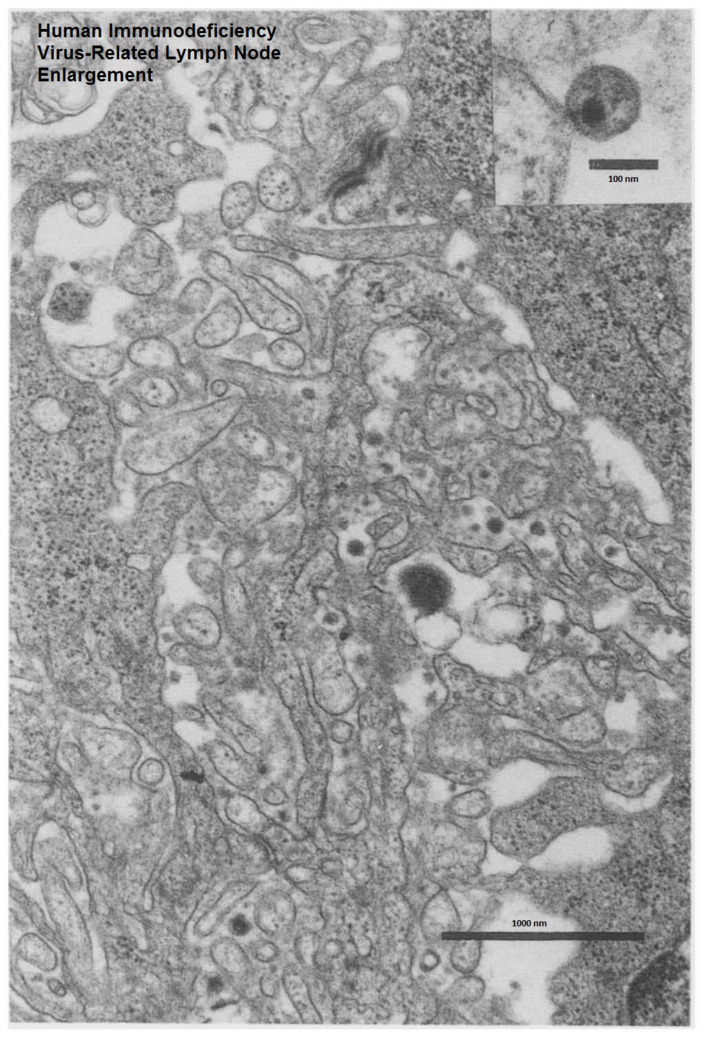
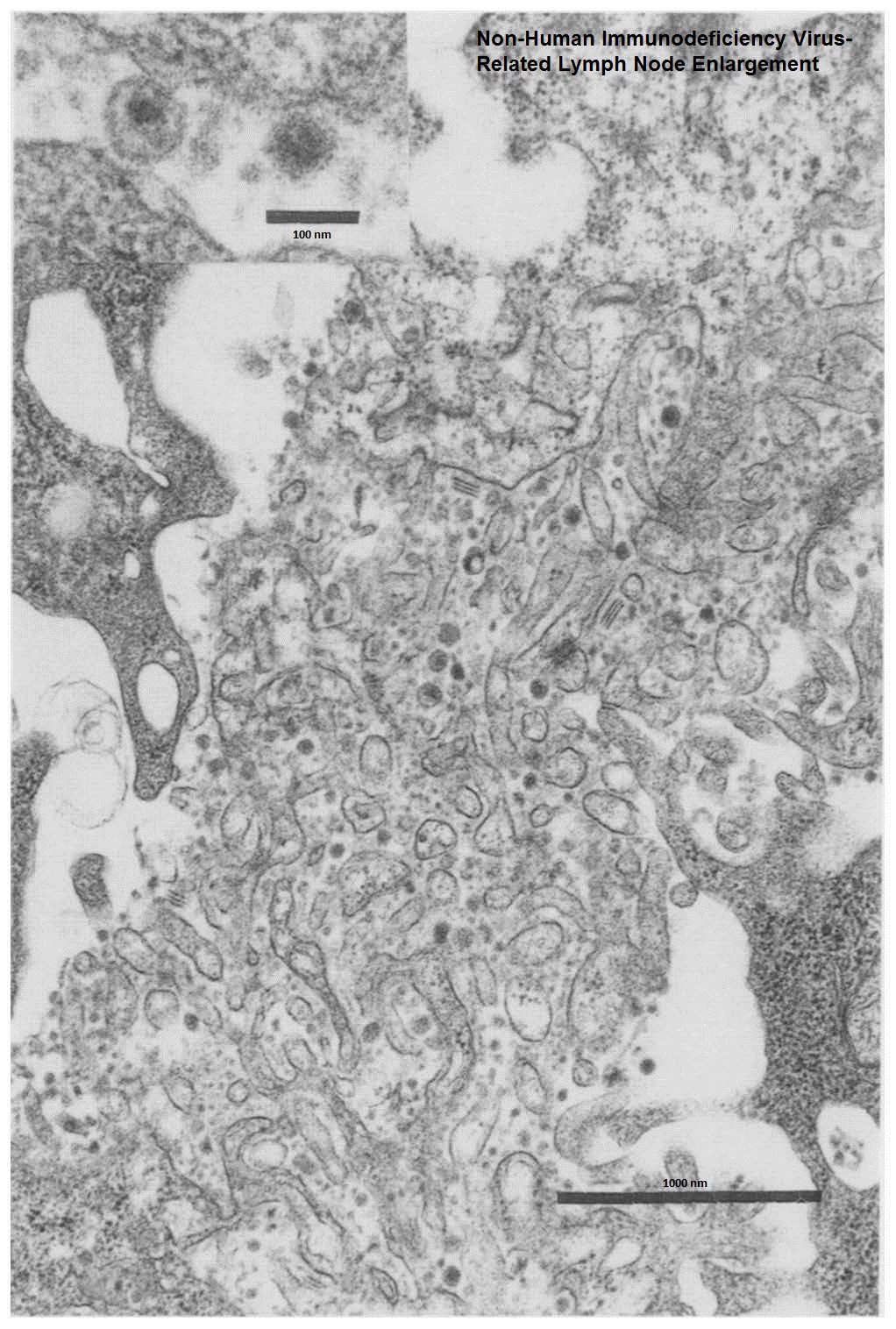
There are dozens of picture of HIV. If these are not a retrovirus what have we been looking at all these years? At its inimical website "Focus on the HIV/AIDS Connection" the National Institutes of Allergy and Infectious Diseases present their evidence "Why is there overwhelming scientific consensus that HIV causes AIDS?". The answer to this question of "consensus" includes an invitation to visit http://www.virology.net/Big_Virology/BVretro.html in order to see "Electron micrographs and other images of HIV". Of these 25 images the majority are diagrams, artists' renditions or computer graphics. Only eight images are electron microscopic pictures and none identify the source or nature of the material photographed. Significantly, none have a size bar, that is, in no EM is it possible to measure the size of the particles or determine the dimensions of any other morphological feature. From the scientific point of view this is both highly unprofessional and unsatisfactory because the dimension of particles is critical to taxonomy. The best conclusion one can draw from the EMs (the other images are irrelevant) is the existence in unidentified cell cultures of minute quantities of matter, that is, particles of indeterminate size which also possess certain other morphological features. Since these are the pictures "we have been looking at all these years", let us examine the proposition "These EMs are a retrovirus HIV". First, how do we interpret this image? Compare it to this EM at the Virology link above . Both EMs show particles "budding" from the cell membrane but, whereas the second image is said to be "HIV", the first is a typical type-C retroviral particle found in a healthy, human placenta [1]. Such particles are present in nearly all human placentas [2,3], a fact scientists have known for at least forty years. Given that when Montagnier discovered HIV he reported "This virus is a typical type-C RNA tumor virus" (tumor virus is the previous name for retroviruses) and Gallo reported his EM as proof his "new isolates are members of the HTLV family", a particular species of type-C particles, do we conclude that nearly all healthy, pregnant women are also infected with a retrovirus HIV? Obviously not but equally as obvious it is essential to exercise great care when interpreting EM images bearing particles with the appearances of retroviruses. We need a definition of a retrovirus particle. Professor Hans Gelderblom from the Koch Institute in Berlin, the world's leading figure in the electron microscopy of "HIV", defines retroviruses as "enveloped viruses with a diameter of 100-120 nm [nanometre=10-9 metre] budding at cellular membranes. Cell released virions contain condensed inner bodies (cores) and are studded with projections (spikes, knobs)" [4]. We also need to know that retroviruses are divided into three subfamilies (0ncovirinae, Lentivirinae and Spumavirinae). The subfamily Oncoviruses are subdivided into the genuses type -A, -B, -C, and -D particles [5]. See here. Retroviral particles also share a physical property of concentrating (banding) at a density of 1.16 gm/ml when centrifuged at high speeds in sucrose density gradients, a fact long used in their purification. See question x for further discussion. When Juliet mused about her lover being a Montague and not a Capulet she said "What�s in a name? that which we call a rose By any other name would smell as sweet". In other words, Juliet was talking about one species of plant. If we are searching for a retrovirus particle in EMs of the tissues of AIDS patients should we expect the same? If HIV were a flower awaiting discovery would we expect it to be a rose, a violet and a geranium all at the same time? We would not yet this is the case when we come back to examining EMs of tissues of AIDS patients. Montagnier and his colleagues reported HIV initially as a type C particle, then as a type D particle and then as a Lentivirus. In 1984 Gallo and his colleagues reported HIV as a type C particle. However, in 1985 Gallo wrote: "A possible unique feature of the virions is the cylindrical core observed in many presumably mature virions. Virions having this type of core have been frequently reported for certain type D retroviruses, and in some instances, for type C retroviruses". Jay Levy reported HIV as a type D particle. Others at the University of California wrote that "AIDS virus isolated show morphologic characteristics of type C, type D and Lentiviruses". According to Anthony Fauci and others" "T-cells and macrophages handle the virus very differently. In the T-cell, virus buds out of the external plasma membrane of the cell. In the monocyte/macrophage cultures it buds into membrane-bound vesicles inside the cells". The latter is a description of a type A, retroviral particle. Thus the leading HIV experts have described HIV as a member of two subfamilies and three genera of Retroviridae. These taxonomical differences imply that if HIV was a newly discovered mammal, it could have been human, a gorilla or an orang-utan. By consensus at present HIV is regarded as a Lentivirus. This agreement was reached when it was realised that in "HIV" positive individuals AIDS did not appear soon after "infection", although Lentivirus is a description based on appearances, not behaviour. But if "HIV" is now a Lentivirus then what Montagnier and Gallo discovered could not have been HIV. How can these and other scientists still claim HIV was discovered in 1983/84 and these papers provide "clearcut" evidence HIV is the cause of AIDS? Thus we see there is more to this problem than meets the eye. The important message is that no amount of searching EMs can prove that a particular particle, even if it looks identical to a retroviral particle, proves the particle is a retrovirus. This follows because the crucial defining property of a virus or retrovirus is its ability to produce identical progeny when introduced into uninfected cell cultures. However, unlike all "other" viruses, retroviruses may appear in cultures in which no external ("exogenous") infection has taken place. That is, they may appear spontaneously, from "within", that is, "endogenously". Retrovirologists claim these retroviruses appear "out of nowhere" because cells contain pre-existing retroviral genetic information passed on from one's parents (known as "vertical" transmission). In fact animal data show that the yield and rate of appearance of such retroviruses can be accelerated a millionfold by chemically stimulating the cell cultures in the same manner as cultures derived from tissues of AIDS patients are manipulated to obtain "HIV". (Significantly, no "HIV" appears in cell cultures unless the cells are manipulated in this manner). Thus to prove the particles claimed to be "HIV" are a retrovirus and not inert, microscopic matter the "HIV" particles must be purified, that is, separated from all other biological material which might also be capable of stimulating the cells to produce similar particles and then added to fresh, "uninfected" cell cultures. If identical particles are obtained and also possess all the morphological and biochemical properties of Gelderblom's definition then a scientist can claim the particles are a retrovirus. In fact this was the approved and publicised method for retroviral research used in the decade before the AIDS era including by present day HIV protagonists such as Barre-Sinoussi and Chermann. If this procedure is not strictly followed then it is impossible to exclude the possibility the particles are the result of being sick with AIDS, or the culture conditions, or both. To date there is no experimental data proving that "HIV" particles have been purified much less data that the same particles generate identical particles. Thus there is no basis for the NIAID claim that the particles seen at the link on their website are a retrovirus. Furthermore, 1. As Hans Gelderblom and his colleagues pointed out in 1998, to date nobody has reported the presence of "infectious plasma HIV". 2. At least a decade before the AIDS era retrovirologists including Gallo knew that particles with appearances similar to retroviruses may be found in cell cultures which had not been "infected" with a retrovirus [6]. These can be than "cellular fragments", microsomes from disrupted cells, "membraneous vesicles which may enclose other cellular constituents including nucleic acids", especially when "inadvertent lysis of cells" was induced. (In "HIV" research lysis is deliberately performed to obtain the "HIV" RNA and proteins). None of these retroviral-like particles are retroviruses because they are inert, that is, they are not replication competent. 3. In no HIV "infected" cultures to date are there particles which fulfil the Gelderblom definition. That is, that display both principle morphological characteristics of retroviruses, that is "a diameter of 100-120nm" AND surfaces which "are studded with projections (spikes, knobs)". 4. In the only EM study, either in vivo or in vitro, in which suitable controls were used and in which extensive blind examination of controls and test material was performed virus particles indistinguishable from "HIV" were foundin 18/20 (90%) of AIDS as well as in 13/15 (88%) of non-AIDS or no risk of AIDS related lymph node enlargements.This led the authors to conclude: "The presence of such particles do not, by themselves indicate infection with HIV" [7]. Seehere andhere. Can you distinguish the AIDS from the non-AIDS patient? Thus the answer to the question "What have we been looking at all these years" should be rephrased "What have we been looking at and not looking at all these years?" [11]. Whatever the answer may be it is presumptuous to claim it is a retrovirus. ENDNOTES (additional references are in the Perth Group papers).
1. Panem S. C Type Virus Expression in the Placenta. Current Topics in Pathology 1979;66:175-189. 2. Retroviral-like particles are ubiquitous [8]. In the 1970s such particles were frequently observed in human leukaemia tissues, cultures of embryonic tissues and "in the majority if not all, human placentas" . Type-C retroviral particles are present in "fish, snakes, worms, pheasant, quail, partridge, turkey, tree-mouse and agouti" as well as in "tapeworms, insects...and mammals"
3. Not only are the same particles as the "original HIV" particle present in the normal, "non-HIV" infected human placenta, so are the "specific" "HIV" proteins p18, p24 and gp120 [9]. Even more surprising, no "HIV" proteins could be found in the placentas of 75 HIV seropositive women [10].
4. Gelderblom HR, �zel M, Hausmann EHS, Winkel T, Pauli G, Koch MA. Fine Structure of Human Immunodeficiency Virus (HIV), Immunolocalization of Structural Proteins and Virus-Cell Relation. Micron Microscopica 1988;19:41-60. 5. Frank H. Retroviridae. In: Nermut MV, Steven AC, editors. Animal Virus and Structure. Oxford: Elsevier; 1987. p. 253-256. 6. Gallo RC, Wong-Staal F, Reitz M, Gallagher RE, Miller N, Gillespie DH. Some evidence for infectious type-C virus in humans. In: Balimore D, Huang AS, Fox CF, editors. Animal Virology. New York: Academic Press Inc.; 1976. p. 385-405. 7. O'Hara CJ, Groopman JE, Federman M. The ultrastructural and immunohistochemical demonstration of viral particles in lymph nodes from human immunodeficiency virus-related and non-human immunodeficiency virus-related lymphadenopathy syndromes. Human Pathology 1988;19(5):545-9. 8. Grafe A. A history of experimental virology. Heidelberg: Springer-Verlag; 1991. 9. Faulk WP, Labarrere CA. HIV proteins in normal human placentae. American Journal of Reproductive Immunolgy 1991;25(3):99-104. 10. Peuchmaur M, Delfraissy JF, Pons JC, Emilie D, Vazeux R, Rouzioux C, et al. HIV proteins absent from placentas of 75 HIV-1-positive women studied by immunohistochemistry. Aids 1991;5(6):741-5. 11. "What have we not been looking at all these years?" or "Why have the CDC, NIAID and other experts failed to publicise the whole "particle" story? If Juliet had decided to present Romeo with a rose to symbolise her love she may well have sent her nurse to pick one from the garden. Let us suppose the nurse could distinguish flowers from trees but was not otherwise horticulturally gifted. She would have no problem bringing back a rose if she were directed to a rose garden where roses were the only plants growing. But if she were directed to a garden display containing several different species of flowers what could she do? She may have brought back a rose but she may have not. The same problem arises in relation to EM data of tissues and cultures from AIDS patients, although the NIAID do not publish these data. "HIV infected cultures" contain in addition to the particles with the morphology attributed to HIV many other "viral particles". For example: Hockley and his colleagues from the Electron Microscopy and Photography Section and Division of Virology at the National Institute for Biological Standards and Control in the United Kingdom describe a profusion of particles which they divide broadly into three groups, mature, ring-like and small with spikes. The mature particles "were approximately spherical in shape and 100 to 150nm in diameter. The outer lipid membrane was frequently broken or absent in places, and there was no evidence of surface spikes...A few mature particles were found that were larger than average and appeared to contain a double nucleoid...in the preparation of HIV there were always many vesicles with granular contents in which it was not possible to recognise a distinct nucleoid". Also, "The ring-like particles had a more consistently spherical shape and were larger (140nm in diameter)" and the small particles "were unusually spherical but sometimes slightly angular in shape and 65 to 90nm in diameter" and had spike-like projections on their surface. Hans Gelderblom who has done most of the EM studies in HIV/AIDS research reported that although Lentiviruses and thus HIV is considered to have a cone shaped core, he and his colleagues found centrosymmetric and tubular cores as well. The caption to one photograph reads: "Virions can be seen having either elongated, 'baton-like' tubular cores 30-35nm in diameter or containing more than one core. Tubular and regular cone-shaped cores can coexist within one virion". The text states: "Rarely, tubular core structures reminiscent of batons with a diameter of 30-35nm and a length of 150-250nm are observed". Lekatsas and other virologists from Pretoria and Johannesburg reported: "We used the characteristic cylindrical structure in the core as an identifying characteristic for the virus to distinguish it from cellular debris and also noted that it may vary considerably in its dimensions and morphological features. We have found two basic virus particle sizes, 90nm and 120nm, both present in large numbers. The larger particle bears no surface projections while the smaller particle is rarely 'naked' and usually bears projections". The CDC reported: HIV particles are "usually round and have a diameter of about 85-95nm...Virus with bar-shaped nucleoids and particles with a tear-drop shape are commonly seen in HTLV-III/LAV infected lymphocytes, sometimes ring-shaped particles without dense nucleoids are also seen". The question then arises if the particles with the "unique" morphology considered to be HIV represent an exogenous retrovirus originating from tissues of AIDS patients or those at risk, then what is the origin and role of the many non-HIV particles and which, if any, of the "HIV" or non-HIV particles band at 1.16 gm/ml? That is, which have the density characteristic of retroviruses? (see question x). Given the profusion of particles that are present in these cultures how does anyone know which particle is "HIV" or which particles, "HIV" or "not-HIV", causes AIDS? The problem is confounded even further because retrovirus-like particles have been found in non-HIV-infected cord blood lymphocytes cell cultures and in other cells used for HIV "isolation" such as and H9 (HUT-78), CEM, C8166 and EBV transformed B-cells. |